Identifying deteriorating patients is the foundation of successful early intervention and rapid response. When clinicians recognize warning signs in a patient and respond promptly, they are better positioned to prevent life-threatening emergencies like cardiac arrest, severe sepsis, and more. But how is it done? One way is by observing, measuring, and tracking a patient’s physiological parameters, known as patient monitoring.
In this article, we’ll cover the basics first: How is patient monitoring different from surveillance, and why is it important? Next, we’ll discuss the top 3 mistakes clinicians make when using patient monitoring devices — and how to avoid them.
Patient monitoring 101
What is patient monitoring?
The terms “monitoring” and “surveillance” are often used interchangeably in clinical settings but are not the same.
Monitoring is the observation, measurement, and recording of physiological parameters. For example, it might look like a clinician taking a patient’s vital signs every 4-6 hours.
On the other hand, surveillance is a systematic, goal-directed process. It includes early detection of changes, interpretation of the clinical implications of those changes, and rapid initiation of appropriate interventions.
In short: Monitoring is an important piece of the larger surveillance process. In surveillance, clinicians take the data collected during patient monitoring and use it to make decisions and intervene as needed.
Why is it important?
Put simply, accuracy matters. Surveillance is one of the primary ways clinicians are able to observe, track, and respond to trends in a patient’s health. But they can’t do that accurately if the data collected during patient monitoring isn’t correct to begin with.
Unfortunately, there are several common mistakes clinicians make when using monitoring devices on patients, either due to time constraints or lack of awareness of the correct method. In the next sections, we’ll talk about what those mistakes are and offer practical tips to avoid the pitfalls.
1. Improper manual blood pressure technique
Overview
When it comes to manual blood pressure measurement, practices and techniques vary considerably across healthcare practitioners. As a result of this inconsistency and lack of standardization, automated blood pressure measurement is the preferred method.1 That said, manual measurement is still used, and there are ways to improve technique and accuracy with this approach.
Tips to improve accuracy
- Use an appropriately sized blood pressure cuff for the patient.
- Place the cuff 2-3 cm above the elbow crease and at heart level.
- Before measurement, have the patient empty their bladder, rest for 5 minutes, and uncross their legs.
- During measurement, make sure that the patient is not speaking.
2. Incorrect telemetry electrode placement
Overview
Telemetry monitoring is used for patients that have (or may have) a concerning arrhythmia. But in order to correctly identify those arrhythmias, accurate electrode placement is a must.2 As an added benefit, correct placement can also decrease false alarms, which is one of The Joint’s Commission’s National Safety Goals.
Unfortunately, just like manual blood pressure technique, electrode placement is another area where there is substantial variation across clinicians. To help address this, the American Heart Association published standards in 2004 and updated them in 2017. Clinicians can (and should) look to these standards for guidance. We’ve also included a few tips below.
Tips to improve accuracy
- Ensure all clinicians are trained and knowledgeable in proper electrode placement and why it matters. Education is crucial.
- Wash and dry the patient’s skin. Do not use alcohol, which can degrade the leads.
- Rub the patient’s skin with gauze to increase capillary blood flow and remove skin cells and oil. This increases the ability to collect and transmit electrical signals.
- Replace electrodes if they are falling off. Allow skin to fully dry before replacing (e.g., if the patient is sweating).
- Check battery levels/switch batteries to decrease false alarms.
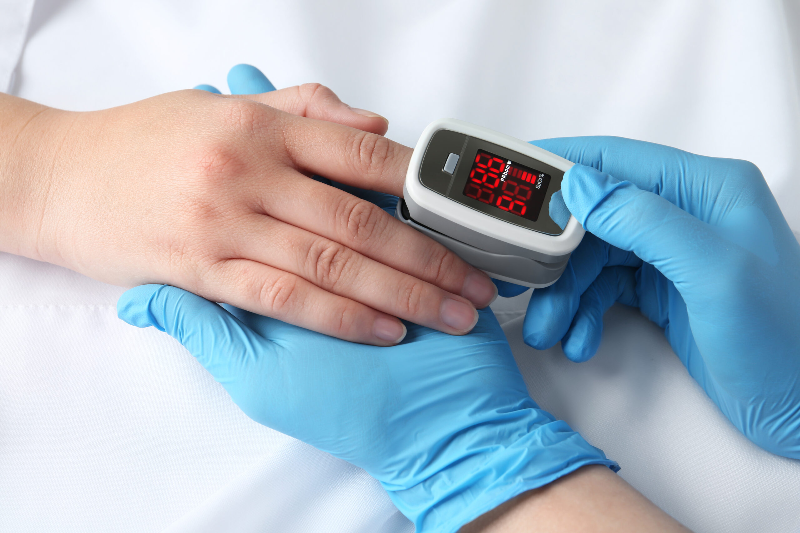
3. Inaccurate pulse oximetry readings
Overview
For a patient with a pulse oximetry reading of 80 to 100%, accuracy is approximately 2% in either direction. Accuracy declines rapidly for readings under 80%. Follow the tips below to improve the likelihood of obtaining a correct reading.
Tips to improve accuracy
- Use proper sensors: A finger probe will not be accurate on the forehead, just as a toe sensor will not be ideal on an earlobe. Be sure to use the correct sensor.
- Prevent interference: The reading may be affected by bright lights directly on the sensor, various probes next to each other, excessive movement by the patient (e.g., shivering), and nail polish. All these barriers should be prevented, when possible.
- Ensure perfusion: Make sure the patient has a blood pressure that is able to reach the extremities. Also check that the probe is not placed on the same side as a continuous blood pressure cuff.3
The bottom line
Details matter. Although optimizing practices around monitoring devices may not seem like a top priority in the grand scheme of patient care, it is an essential piece of patient surveillance. Done correctly, it helps clinicians identify deteriorating patients and make accurate decisions. Ensuring correct use of monitoring devices optimizes patient care, promotes health and safety, and prevents harm.
RELATED ARTICLES


Keep learning
Patient monitoring & surveillance are at the core of successful rapid response systems. Now that you’ve mastered the basics of patient monitoring, read our next blog article for a closer analysis of how rapid response teams benefit patients and staff.
References
-
Choudhury, A., & Asan, O. (2020). Role of artificial intelligence in patient safety outcomes: Systematic literature review. JMIR Medical Informatics, 8(7), e18599. https://doi.org/10.2196/18599
-
Medani, S. A., Hensey, M., Caples, N., & Owens, P. (2018). Accuracy in precordial ECG lead placement: Improving performance through a peer-led educational intervention. Journal of Electrocardiology, 51(1), 50–54. https://doi.org/10.1016/j.jelectrocard.2017.04.018
-
Vos, J. J., Ellermann, S. F., & Scheeren, T. L. (2019). Journal of clinical monitoring and computing 2017/2018 end of year summary: Monitoring—and provocation—of the microcirculation and tissue oxygenation. Journal of Clinical Monitoring and Computing, 33(2), 201–209. https://doi.org/10.1007/s10877-019-00270-7